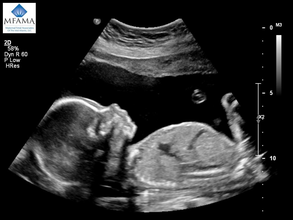
The scan will go as following. An experienced ultrasound technician will be the one conducting the ultrasound.
 Maternal Fetal Associates Of The Mid Atlantic Outstanding Care For You And Your Baby
A high-risk obstetric patient is one who has the potential for or is at an increased risk for an adverse maternal or fetal pregnancy outcome.
High risk pregnancy ultrasound. These complications will require special attention from your doctor. Pediatric medical and surgical specialists advise you during pregnancy and treat your baby during childhood. No one wants their pregnancy to be high risk but if it is this is a wonderful place to go.
At its extreme adverse would mean maternal or fetal injury or death. Our ultrasound experts called sonographers focus exclusively on obstetrical and gynecological ultrasounds performing more than 5000 tests every year. High risk is defined by a diagnosis code from the UnitedHealthcare Community Plan Medicaid ICD-10-CM Detailed and High Risk Fetal Ultrasound Diagnosis list.
Typically done between weeks 10 and 12 of pregnancy CVS can identify certain genetic conditions. These include high temperatures radiation exposure and high altitudes. Your first ultrasound typically occurs around the 12th week of pregnancy.
This noninvasive imaging test is helpful for routine and high-risk pregnancies. High Risk Pregnancy Fetal Medicine Specialists OBGYNs located in Ventura CA Thousand Oaks CA West Hills CA and Mission Hills CA. When identifying the high-risk pregnant patient both the mother and the fetus must be considered.
Ultrasound can be a routine test but it can also be a high risk test when the doctor is evaluating a specific high-risk issue such as pre-eclampsia intrauterine growth restriction placenta previa and so forth. Your health care provider might use an ultrasound to measure the length of your cervix at prenatal appointments to determine if youre at. This test can detect approximately 90 to 92 percent of fetuses with Down syndrome and 90 percent of fetuses with Trisomy 18 while maintaining a false positive rate of 5 percent.
HIGH RISK OBSTETRIC ULTRASOUND DICTATION GUIDELINES NEURO CHOROIDAL SEPARATION Choroidal separation in the region of the atrium greater than or equal to 4mm recommend follow up in 6 weeks to re-evaluate making sure to note which ventricle is abnormal and attempting to see both ventricles. Our Division of Maternal-Fetal Medicine also known as High-Risk Obstetrics provides expert multidisciplinary care for women and newborns who have complications identified prior to or during pregnancy. Here are some factors that make for a high-risk pregnancy.
This provides women at the highest risk for having a baby with Down syndrome or Trisomy 18 with results in the first trimester. Prenatal ultrasounds are something to look forward to because they provide your first glimpse of your baby. A High Risk Pregnancy will be very similar to the first and second trimester ultrasounds you may have had already.
High-Risk Pregnancy Care at Brigham and Womens Hospital. Sometimes diabetic patients will have cardiac anomalies affecting their fetus and this can also be monitored with ultrasound. What is the Procedure of a High Risk Pregnancy Ultrasound.
Ultrasound for cervical length. Conveniently located for families in the Snohomish and Skagit Counties the Maternal Fetal Medicine Clinic at Arlington offers highly specialized care for women with health conditions or special challenges that may complicate pregnancy. Maternal-fetal medicine specialists from the University of Washington consult with us as needed.
However some doctors only conduct this exam if you have certain high-risk. They also allow your OBGYN to examine your developing baby. In 4 reviews Was seen here in Nov 2020 instead of my typical OB because I was dialted at my 20 week ultrasound.
You or your partner have a family history of genetic concerns. The significance of Doppler ultrasound in evaluating pregnancies that have the risk for preeclampsia intrauterine growth restriction fetal anaemia and umbilical cord abnormalities has become indispensable. High-risk obstetricians check the health of both mother and baby during pregnancy.
A pregnancy is considered high-risk when there are potential complications that could affect you your baby or both. Understand ultrasound services at Swedish Maternal and Fetal Specialty Center. Discuss the role of sonography in the high-risk pregnancy.
Unless a different limit is outlined by the State claims for the fourth andor subsequent OB ultrasound procedure per pregnancy must contain a high risk pregnancy diagnosis code. Your first ultrasound also known as a sonogram may take place when youre around 6 to 8 weeks pregnant. Specialized care for expecting mothers with high-risk pregnancies.
Follow protocol for history of hydrocephalus. Recent findings aided in timing delivery of severely growth-restricted fetuses by promoting the use of ductus venosus Doppler.

/1745246_color1-5b9fd36cc9e77c002ce2dafa.png)
