The person who performs an ultrasound scan is called a sonographer but the images are interpreted by radiologists cardiologists or other specialists. A chest ultrasound is a noninvasive diagnostic exam that produces images which used to assess the organs and structures within the chest such as the lungs mediastinum area in the chest containing the heart aorta trachea esophagus thymus and lymph nodes and pleural space space between the lungs and the interior wall of the chest.
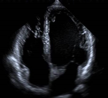 Heart Ultrasound Echocardiogram Insight Medical Imaging
It enables physicians to view.
/1745246_color1-5b9fd36cc9e77c002ce2dafa.png)
Ultrasound of the heart is called. Cardiac ultrasound is also referred to as echocardiogram cardiac echo or transthoracic echo TTE. Ann Reed the person that performs our abdominal ultrasounds also does echocardiograms. The device sends special sound waves called ultrasound through your chest wall to your heart.
This non-invasive procedure provides information about the size and shape of the heart its pumping capacity and blood flow through the heart chambers. An ultrasound scan is routine for pregnant women. Detect abnormalities of heart structures such as the heart valves.
See the separate leaflet called Echocardiogram for more details. Also known as an echocardiogram or ECHO this type of medical imaging uses a wand-like transducer or probe that emits high-frequency sound waves to create an image of your heart. Stress echo is done as part of a stress test.
An echocardiogram echo is a graphic outline of the hearts movement. Ultrasonic images also known as sonograms are made by sending pulses of ultrasound into tissue using a probe. It uses ultrasonic waves that bounce off the heart to create a moving image of it.
Blockages to blood flow such as clots. An ultrasound of the heart is called an echocardiogram. The ultrasound pulses echo off tissues with different reflection properties and are recorded and displayed as an image.
The human ear cant hear ultrasound waves. As the ultrasound waves bounce off the structures of your heart a computer in the echo machine converts them into pictures on a screen. An echocardiogram echo is a test that uses high frequency sound waves ultrasound to make pictures of your heart.
Heart ultrasound is known as an echocardiogram which is a sonogram of the heart and can be called cardiac ultrasound or cardiology ultrasound. For symptoms like shortness of breath heart palpitations or chest pain your doctor might request a heart ultrasound to help find the cause for your concerns. Fred Brewer our cardiologist for echocardiograms.
Ad All New Patients Welcome at Memorial City Cardiology Associates. Cardiac Ultrasound or Echocardiogram. An echocardiogram or echo is a specialized ultrasound of the heart using 2-D and Doppler ultrasound.
The test is also called echocardiography or diagnostic cardiac ultrasound. Ultrasound of the heart is commonly called an echocardiogram or echo for short. Ad All New Patients Welcome at Memorial City Cardiology Associates.
During a stress test you exercise or. During an echo test ultrasound high-frequency sound waves from a hand-held wand placed on your chest provides pictures of the hearts valves and chambers and helps the sonographer evaluate the pumping action of the heart. This type of ultrasound scan is called echocardiography.
We more commonly utilize the expertise of Dr. Ultrasound are sound waves with frequencies which are higher than those audible to humans 20000 Hz. Doppler ultrasound images can help the physician to see and evaluate.


