Showing posts with label disorders. Show all posts

Saturday, August 21, 2021

X Linked Genetic Disorders

People with this disease often have progressive loss of the fatty covering myelin that surrounds the nerves in the brain and spinal cord. X-linked disorders result from mutated genes on the X chromosome.
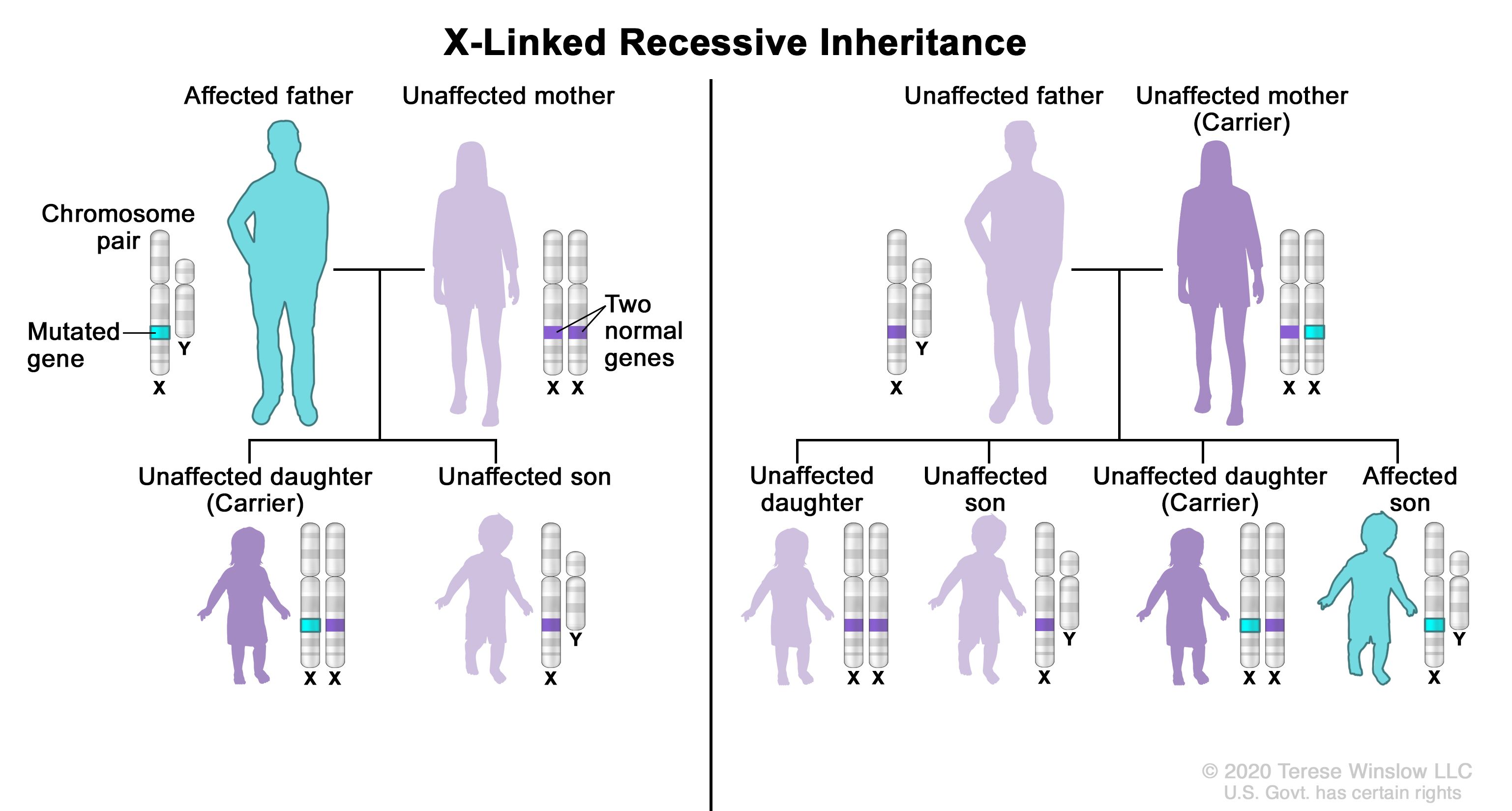
Definition Of X Linked Recessive Inheritance Nci Dictionary Of Genetics Terms National Cancer Institute

Humans and other mammals have two sex chromosomes the X and the Y.

X linked genetic disorders. However the Y chromosome doesnt contain most of the genes of the X chromosome. The scales of X-linked ichthyosis are often dark and usually cover. Females inherit two X chromosomes with males inheriting just one.

Their expression in females and males is not the same. Males have one X and one Y. There are several X-linked or sex-linked recessive genetic disorders hemophilia muscular dystrophy which are inherited through a genetic defect on an X chromosome.

One X chromosome is inherited from each parent XY. Genes on the X chromosome are said to be X-linked. X-linked ichthyosis is a disorder in which the skin cells are produced at a normal rate but they do not separate properly at the surface of the stratum corneum the outermost layer of the skin.

X-linked inheritance means that the gene causing the trait or the disorder is located on the X chromosome. Genes on the X chromosome can be recessive or dominant. Males have one X chromosome.

X-linked adrenoleukodystrophy X-ALD is a genetic disease that affects the nervous system and the adrenal glands small glands located on top of each kidney. There are around 4000 known inherited conditions that are caused by a difference in a single gene and a number of these follow an X-linked pattern of inheritance. X-linked recessive diseases most often occur in males.

On the other hand females who have two X chromosomes will be carriers of the defect in the majority of cases and so they are usually asymptomatic. An X-linked genetic disorder is one caused by a mutation change in a gene on the X chromosome. This represents the sex chromosomes of a female.

A male has an X chromosome from his mother and a Y chromosome from his father. Cell-cloned pigs from male cells with mutations in an X chromosomal gene is a reliable and straightforward method for reproducing X-linked genetic diseases XLGDs in pigs. The three best-known disorders which seem to be inherited in this way are incontinentia pigmenti IP Bloch-Sulzberger oral-facial-digital I OFD I syndrome and focal.

The key difference between X linked dominant and X linked recessive is that X linked dominant is a genetic disorder caused due to a dominant mutant gene located on the X chromosome while X linked recessive is a genetic disorder caused due to one or two recessive mutant genes located on the X chromosomes. It causes progressive muscle weakness and starts to affect boys when they are toddlers. Duchenne Muscular Dystrophy DMD is an X-linked recessive condition.

X-linked is a trait where a gene is located on the X chromosome. X-linked genes have distinctive inheritance patterns because they are present in different numbers in females XX and males XY. Describes a gene on the X chromosome.

In an X-linked or sex linked disease it is usually males that are affected because they have a single copy of the X chromosome that carries the mutation. Females have two X chromosomes. However the severe symptoms of XLGDs are often accompanied by impaired growth and reproductive disorders which hinder the reproduction of.

Because of that it doesnt protect the male. X-linked genetic diseases therefore affect males and females differently. Females have two X chromosomes.

Males have no spare X chromosome to revert to if. Patients with XLH have inactivating mutations in the PHEX gene which encodes phosphate-regulating neutral endopeptidase PHEX with many different mutations of. X-linked human genetic disorders are much more common in males than in females due to the X-linked inheritance pattern.

Males have only one X chromosome. The X-linked dominant disorder X-linked hypophosphatemia XLH is the most common form of inherited hypophosphatemia. In fact feasible production of model pigs reproducing XLGDs such as severe combined immunodeficiency SCID 8 9 Duchenne muscular dystrophy DMD 10 and hemophilia 11 has been reported.

Nuclear transfer using male cells carrying mutations in an X chromosomal gene enables the direct cell-to-animal generation of models for X-linked genetic diseases XLGDs. X-linked dominant inheritance with lethality in hemizygous males is a rare mode of inheritance. Males who have only one X chromosome ie they are hemizygous will fully express an X-linked disorder.

The Y chromosome is the other half of the XY gene pair in the male. This slows the rate of shedding of the skin cells resulting in a build-up of scales. A single recessive gene on that X chromosome will cause the disease.

This represents the sex chromosomes of a male. A female has 2 X chromosomes one she inherited from her mother and one she got from her father.

Friday, May 24, 2019

Blood Tests For Clotting Disorders

It is then analysed in the laboratory. Testing may help women weigh the risks of estrogen-containing contraceptives.

Coagulation Disorders Coagulopathy Introduction Coagulation Disorders Hemostasis Management

A blood test can determine whether the persons clotting is abnormally slow.

Blood tests for clotting disorders. To evaluate someone for recurrent blood clots. These tests are identical to many other blood tests and the risks and side effects are least. Women who have an inherited blood clotting disorder have a greater risk of developing a blood clot while using estrogen a hormone normally produced by the body which may also be supplemented.

Genetic testing may be an advantage in the following circumstances. Measures quantity of antithrombin. Other tests can measure the overall coordinated function of the many proteins needed for normal blood clotting clotting factors.

APTT Partial Thromboplastin Time This test is used as a screening test to evaluate bleeding disorders. Do we have any tests to detect it. The levels of individual clotting factors can also be determined.

The levels of individual clotting factors can also be determined. There is a test people can be given after vaccination to see if they have antibodies against platelet factor 4 in their plasma. The most common of these tests are the prothrombin time PT and the partial thromboplastin time PTT.

The levels of individual clotting factors can also be determined. Testing can help your doctor assess your risk of excessive bleeding. Before you undergo invasive procedures and operations if you suffer from blood clotting disorders you should undergo an assessment that covers important information such as clinical features including personal or family history of bleeding medical conditions present and medications taken.

If it is further blood tests to measure the levels of factor VIII and IX can confirm the diagnosis of hemophilia and can determine its type and severity. Blood-clotting disorders are a group of conditions in which there is too much clotting. When the TT is abnormal disseminated intravascular coagulation the presence of plasma heparin or a hepatopathy should be suspected.

The most common of these tests are the prothrombin time PT and the partial thromboplastin time PTT. General blood clotting tests. They are often inherited.

To evaluate recurrent blood clots andor miscarriages. An abnormal PT with or without an abnormal aPTT but with normal results in the other three tests indicates an abnormal reduction in the vitamin K-dependent clotting factors II VII IX X or factor V. Coagulation tests measure your bloods ability to clot and how long it takes to clot.

This test is used to monitor your condition if you are taking heparin. Other tests can measure the overall coordinated function of the many proteins needed for normal blood clotting clotting factors. As a result an expert in blood clotting conditions needs to explain the blood test results.

The levels of individual clotting factors can also be determined. Low activity may increase risk of clots. PT APTT FBC activated protein C resistance APCR Factor V mutation Leiden assay when APCR is abnormal homocysteine cardiolipin antibodies protein C protein S antithrombin and a prothrombin 20210 mutation test.

Fibrin is the major protein that maintains a clot. The most common of these tests are the prothrombin time PT and the partial thromboplastin time PTT. Patients with APLAS who get blood clots need to be treated with blood thinners and should be managed by a doctor who specializes in blood clotting disorders.

Complete blood count CBC. Your INR will help your healthcare provider determine how fast your blood is clotting and whether your warfarin Coumadin dose needs to be changed. Other tests can measure the overall function of the many proteins needed for normal blood clotting clotting factors.

For example the prothrombin time PT and the activated partial thromboplastin time APTT are commonly done. Several substances are added to the sample. To detect acquired or inherited deficiency.

Tests which may be undertaken to assist the haematologist in determining the risk of recurrent blood clots include. This test helps the doctor to assess the risk of developing clots thrombosis or excessive bleeding in blood vessels. Activated partial thromboplastin time aPTT.

Measures the time it takes blood to clot. A blood sample is taken into a bottle that contains a chemical which prevents the blood from clotting. It measures the number of seconds it takes for a fibrin clot to form in a blood sample.

Special blood tests are needed to test for APLAS and the results can be tricky to understand. Other tests can measure the overall function of the many proteins needed for normal blood clotting clotting factors. A coagulation test measures bloods capability to clot and if it clots how long it does take to clot.

There are a number of tests that may be done. Measures the activity of antithrombin. Your doctor may also decide to do some blood tests.

The most common of these tests are the prothrombin time PT and the partial thromboplastin time PTT.

How To Get Rid Of Lice With Vinegar And Listerine

None of the nits fell off the hair strands and I had to drag them down the entire hair strand using two fingers. Keep the Listerine do its ...