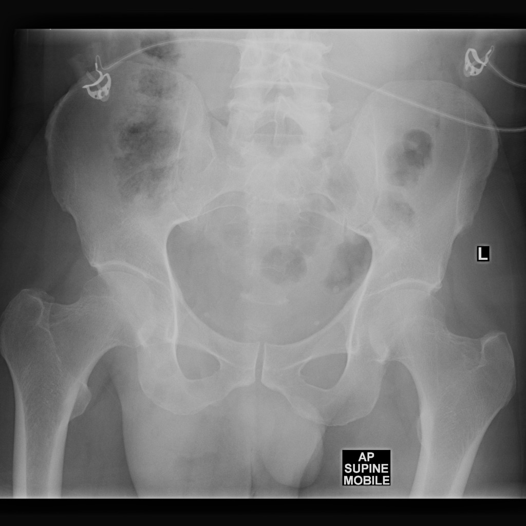
Type III is the 2nd most common type but it is rare compared to type I sliding HH. Inguinal hernia This image shows multiple loops of dilated small bowel indicating small bowel obstruction.
 Sliding Hiatus Hernia Radiology Case Radiopaedia Org
The metallic substance coats the esophagus and stomach helping to isolate them in the.

Hernia x ray. It will classically appear as a mass with an air fluid level behind the heart. The preferred method of diagnosis of a hiatal hernia is an upper gastrointestinal GI barium study. That is because most commonly the stomach herniates into a hiatal hernia.
Most common 95 located posterolaterally. Demographics and etiology Congenital. A hiatal hernia will often be visible on a chest X-ray.
Commonly referred to as a barium swallow the test requires you to drink roughly one-and-a-half cups of chalky fluid containing barium sulfate and about 30 minutes later undergo a series of X-rays. An abdominal X-ray or CT scan may be ordered to look for a hernia and determine if it is strangulated or incarcerated. For this test contrast dye is injected into the muscles through a small needle.
Often a patient with symptoms that are thought to arise from an abnormality in the chest will get a chest X-ray. To diagnose a hiatal hernia your doctor may do tests including. In most cases it can be found by physical exam.
Management and Treatment Do all inguinal hernias require surgery. Diaphragmatic hernias alternative plural. Herniae are defined as either congenital or acquired defects in the diaphragm.
CT scans use X-rays to create images of your abdomen and its organs and they may involve having a contrast dye injected into your arm. Herniation of the stomach above the diaphragm hiatus hernia is a common finding on a chest X-ray Hiatus hernias may be very large as in this image Seeing a gasfluid level helps to make the diagnosis Hiatus hernia - Small. X-rays are then taken to see if you have a hernia.
If there is a hernia hole in the abdominal wall the. A loop of bowel is seen below the level of the inguinal ligament indicating an inguinal hernia the cause of. A liquid that shows on x-rays radio-opaque has to be injected into the abdominal cavity.
There are two main types of congenital diaphragmatic hernia CDHs which are uncommon yet distinct entities that usually occur on the left side 80 of the diaphragm 12. The abdominal X-ray should not be relied on to detect hernias. You drink a liquid that shows up on an X-ray so your doctor can get a better look at your esophagus and.
Herniagram This is a special x-ray not often done now partly because it is invasive that involves an injection with a needle. Type I is a sliding HH and types II-IV are paraesophageal hernias. How to diagnose hiatus hernia on chest x-ray.
Your doctor puts a long thin tube called an endoscope down your throat. When it cannot be found that way you may need a herniogram. Right Esophagram in a patient with type I sliding HH shows the lower esophageal sphincter or phrenic ampulla marked by the A.


/3133026-how-long-before-std-symptoms-appear-071-5aa7dbbb1f4e130037d636a2.png)


:max_bytes(150000):strip_icc()/why-do-i-have-a-headache-after-childbirth-1719586-5c046eeb46e0fb0001c253cf.png)


